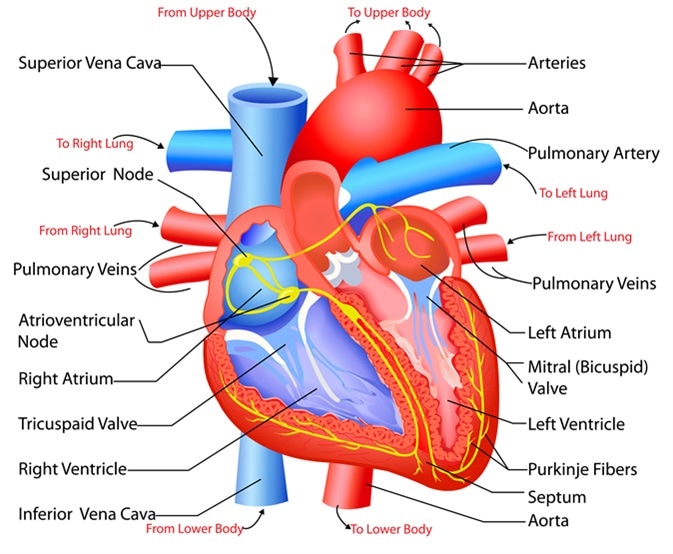
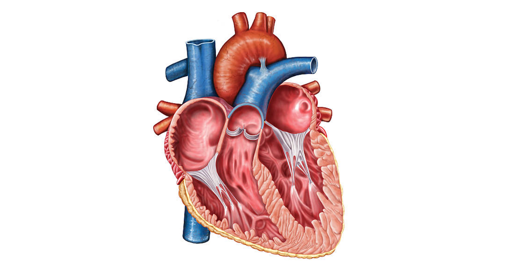
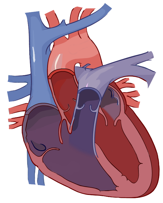
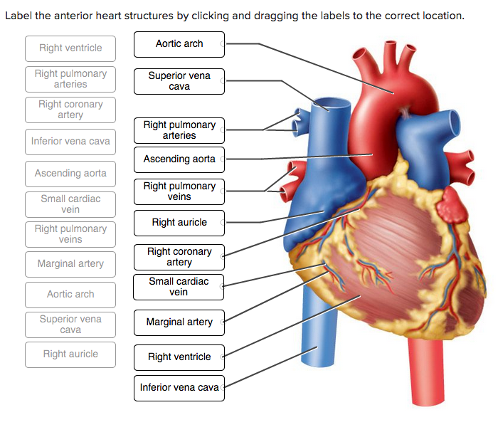
39 heart structure with labels
How to Draw the Internal Structure of the Heart (with Pictures) - wikiHow Coloring and Labeling 1 Color these pink: Border Left Atrium Right Atrium Pulmonary Veins 2 Color these purple: Pulmonary Artery Left Ventricle Right Ventricle 3 Color these blue: Superior Vena Cava Inferior Vena Cava 4 Color this red: Aorta 5 Make sure to label the following: Superior Vena Cava Inferior Vena Cava Pulmonary Artery Pulmonary Veins ebook - Wikipedia An ebook (short for electronic book), also known as an e-book or eBook, is a book publication made available in digital form, consisting of text, images, or both, readable on the flat-panel display of computers or other electronic devices.
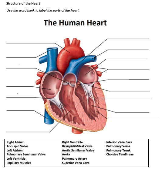
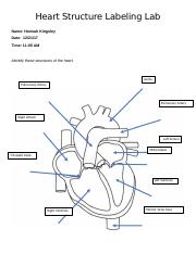
Human Heart - Anatomy, Functions and Facts about Heart - BYJUS Label the Heart Diagram below: Practice your understanding of the heart structure. Drag and drop the correct labels to the boxes with the matching, highlighted structures. Instructions to use: Hover the mouse over one of the empty boxes. One part in the image gets highlighted.
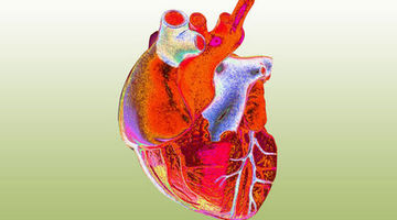
Heart structure with labels
Marketing Week | marketing news, opinion, trends and jobs Marketing Week offers the latest marketing news, opinion, trends, jobs and challenges facing the marketing industry. Heart Diagram with Labels and Detailed Explanation - Collegedunia The heart is located under the ribcage, between the lungs and above the diaphragm. It weighs about 10.5 ounces and is cone shaped in structure. It consists of the following parts: Heart Detailed Diagram. Heart - Chambers There are four chambers of the heart. The upper two chambers are the auricles and the lower two are called ventricles. [IGCSE/GCSE] Heart Structure - Memorize In 5 Minutes Or Less! 📘 FREE Comprehensive notes onhttps:// ️Join patreon to access EXCLUSIVE content! ️ Past ...
Heart structure with labels. U.S. appeals court says CFPB funding is unconstitutional ... Oct 20, 2022 · The structure has been the target of legal challenges before. In this decision, the court ruled in favor of a lawsuit from two trade groups seeking to overturn the CFPB’s 2017 payday lending rule. Because the CFPB’s funding is unconstitutional, the decision said, the rule itself is invalid. Human Heart Diagram Labeled | Science Trends The heart has four different chambers: the left and right ventricles and the left and right atriums. The chambers of the heart and the valves that regulate blood flow to them are considered the plumbing of the heart. The left ventricle and left atrium make up the left heart while the right ventricle and right atrium make up the right heart. Latest Breaking News, Headlines & Updates | National Post Read latest breaking news, updates, and headlines. Get information on latest national and international events & more. Structure of the Heart | SEER Training - National Cancer Institute The outer layer of the heart wall is the epicardium, the middle layer is the myocardium, and the inner layer is the endocardium. Chambers of the Heart The internal cavity of the heart is divided into four chambers: Right atrium Right ventricle Left atrium Left ventricle The two atria are thin-walled chambers that receive blood from the veins.
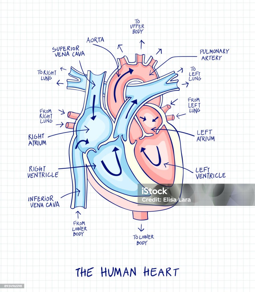
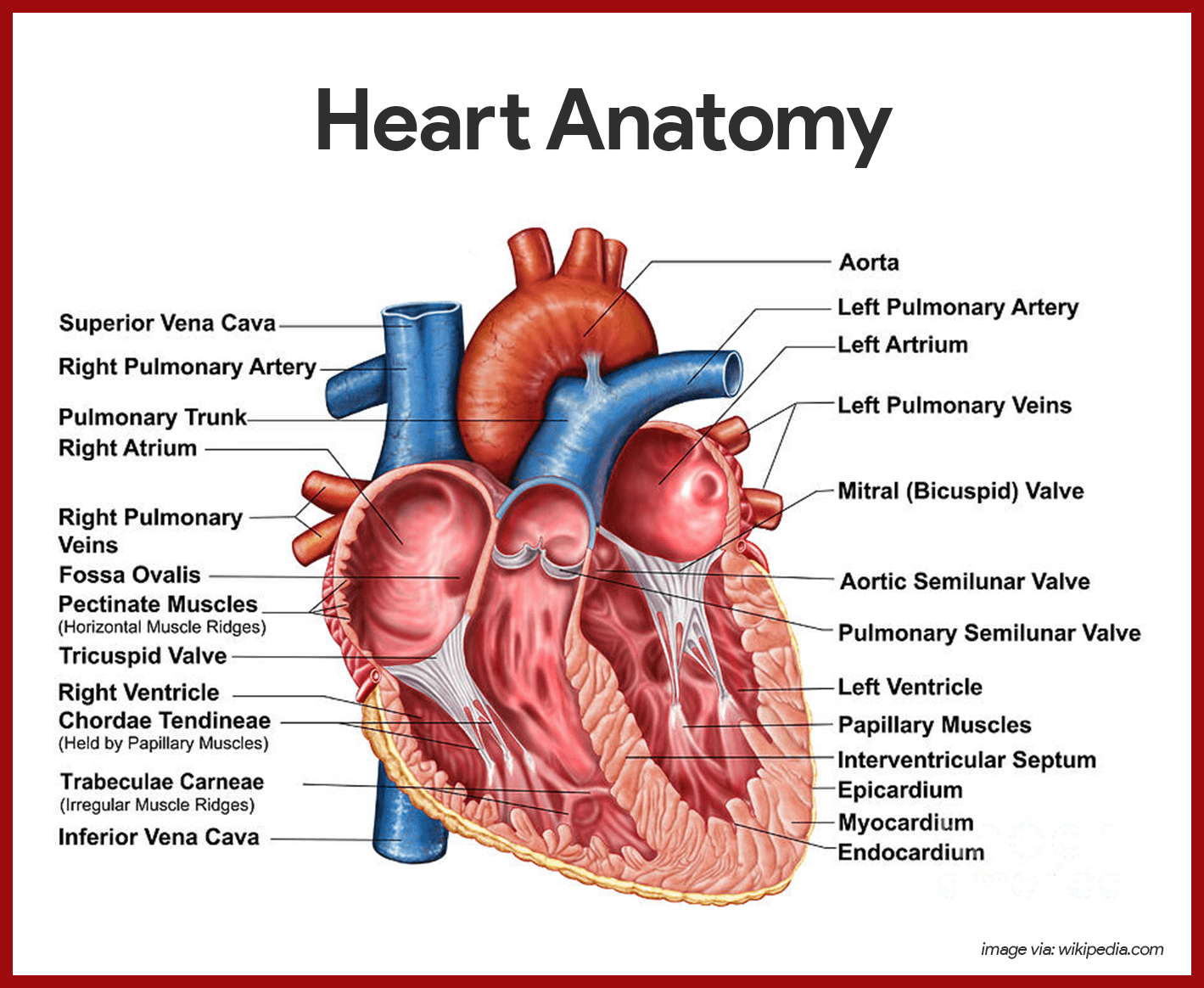
Picture of the Heart - WebMD The heart has four chambers: The right atrium receives blood from the veins and pumps it to the right ventricle. The right ventricle receives blood from the right atrium and pumps it to the... Heart Diagram with Labels and Detailed Explanation - BYJUS Well-Labelled Diagram of Heart The heart is made up of four chambers: The upper two chambers of the heart are called auricles. The lower two chambers of the heart are called ventricles. The heart wall is made up of three layers: The outer layer of the heart wall is called epicardium. The middle layer of the heart wall is called myocardium. Label the Heart Quiz - PurposeGames.com Ummmmmmm . . . it's pretty self explanatory . . . you label the heart. Just remember one thing - you're looking at the heart like it's in someone else so right and left are switched around. This quiz has tags. Click on the tags below to find other quizzes on the same subject. Anatomy. Label the Heart Diagram | Quizlet Label the Heart 4.6 (50 reviews) + − Learn Test Match Created by bluesas9 Terms in this set (15) Superior Vena Cava ... Right Ventricle ... Left Atrium ... Atrioventricular/Tricuspid Valve ... Atrioventricular/Mitral Valve ... Septum ... Right Atrium ... Semi-lunar Valves ... Left Pulmonary Veins ... Right Pulmonary Veins ...
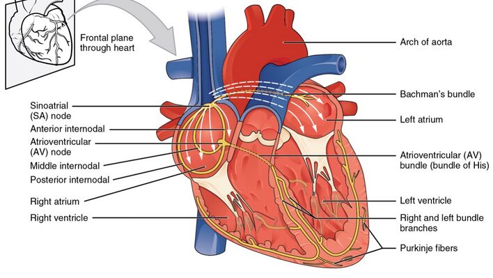
The Anatomy of the Heart, Its Structures, and Functions - ThoughtCo The heart wall consists of three layers: Epicardium: The outer layer of the wall of the heart. Myocardium: The muscular middle layer of the wall of the heart. Endocardium: The inner layer of the heart. Cardiac Conduction Cardiac conduction is the rate at which the heart conducts electrical impulses. Diagram of Human Heart and Blood Circulation in It A heart diagram labeled will provide plenty of information about the structure of your heart, including the wall of your heart. The wall of the heart has three different layers, such as the Myocardium, the Epicardium, and the Endocardium. Here's more about these three layers. Epicardium Health News | Latest Medical, Nutrition, Fitness News - ABC ... Nov 01, 2022 · Get the latest health news, diet & fitness information, medical research, health care trends and health issues that affect you and your family on ABCNews.com Heart Anatomy Labeling Game - PurposeGames.com This is an online quiz called Heart Anatomy Labeling Game. There is a printable worksheet available for download here so you can take the quiz with pen and paper. Your Skills & Rank. Total Points. 0. Get started! Today's Rank--0. Today 's Points. One of us! Game Points. 19. You need to get 100% to score the 19 points available.
Free Anatomy Quiz - The Anatomy of the Heart - Quiz 1 The circulatory system - lower body image, with blank labels attached; The circulatory system - a PDF file of the upper and lower body for printing out to use off-line; Articles: Describe and explain the function of the circulatory system - The circulatory system consists of the heart, the blood vessels (veins, arteries, and capillaries), and ...
Easy way to draw heart structure by 5 steps | labeling of heart ... My youtube channel : facebook page : way to draw hea...
Anatomy Online - Quiz: Anatomy of The Heart Test prep cardiovascular system: structure of the heart. Free interactive quiz for students biology, anatomy and physiology.
Structure and Function of the Heart - News-Medical.net Structure of the heart. The heart wall is composed of three layers, including the outer epicardium (thin layer), middle myocardium (thick layer), and innermost endocardium (thin layer). The ...
Heart Diagram for Kids - Bodytomy As you can see in the diagram of the heart, that heart is divided in four chambers, namely, right atrium, left atrium, right ventricle and left ventricle. Each of the chambers is separated by a muscle wall known as Septum. The left side of the heart receives oxygen rich blood from the lungs and pumps it out the whole body.
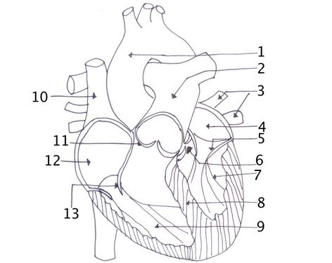
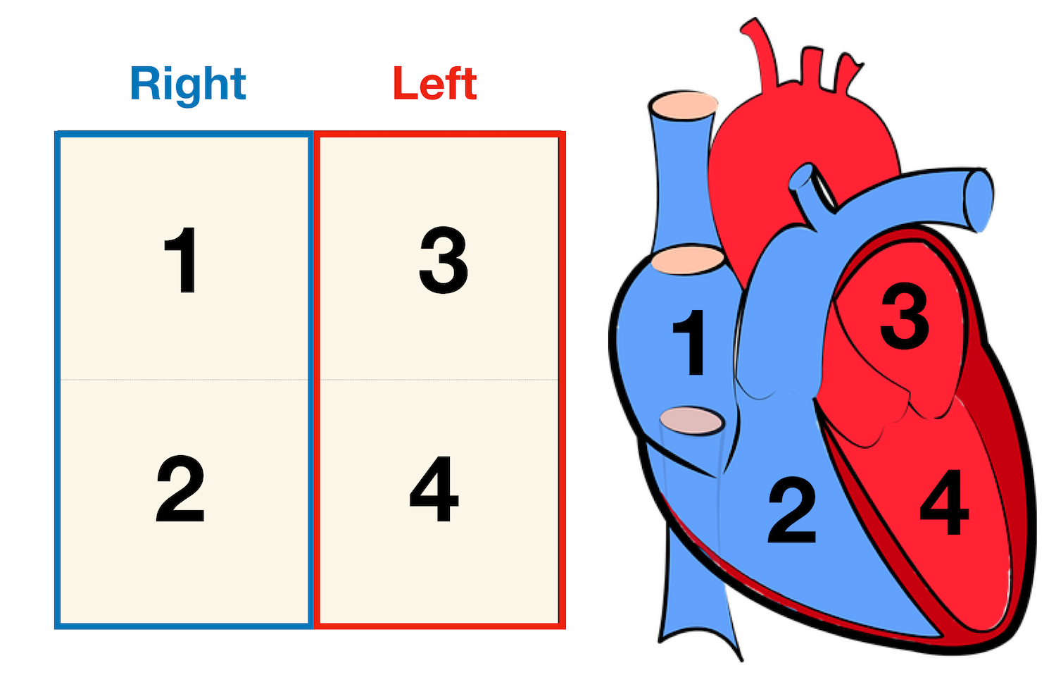
Heart Labeling Quiz: How Much You Know About Heart Labeling? Here is a Heart labeling quiz for you. The human heart is a vital organ for every human. The more healthy your heart is, the longer the chances you have of surviving, so you better take care of it. Take the following quiz to know how much you know about your heart. Questions and Answers 1. What is #1? 2. What is #2? 3. What is #3? 4. What is #4?
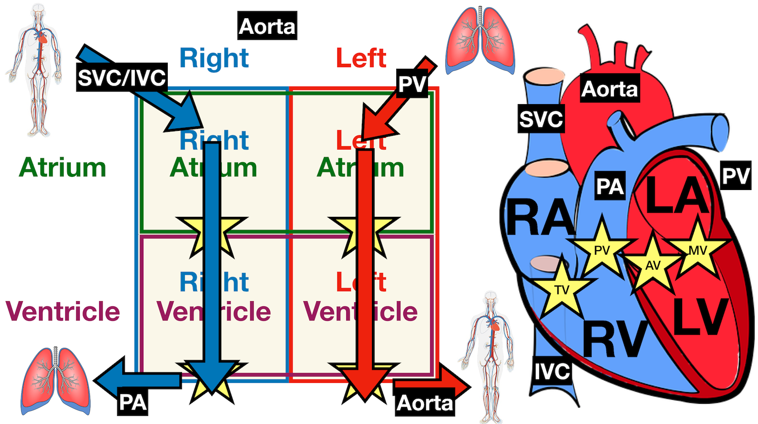
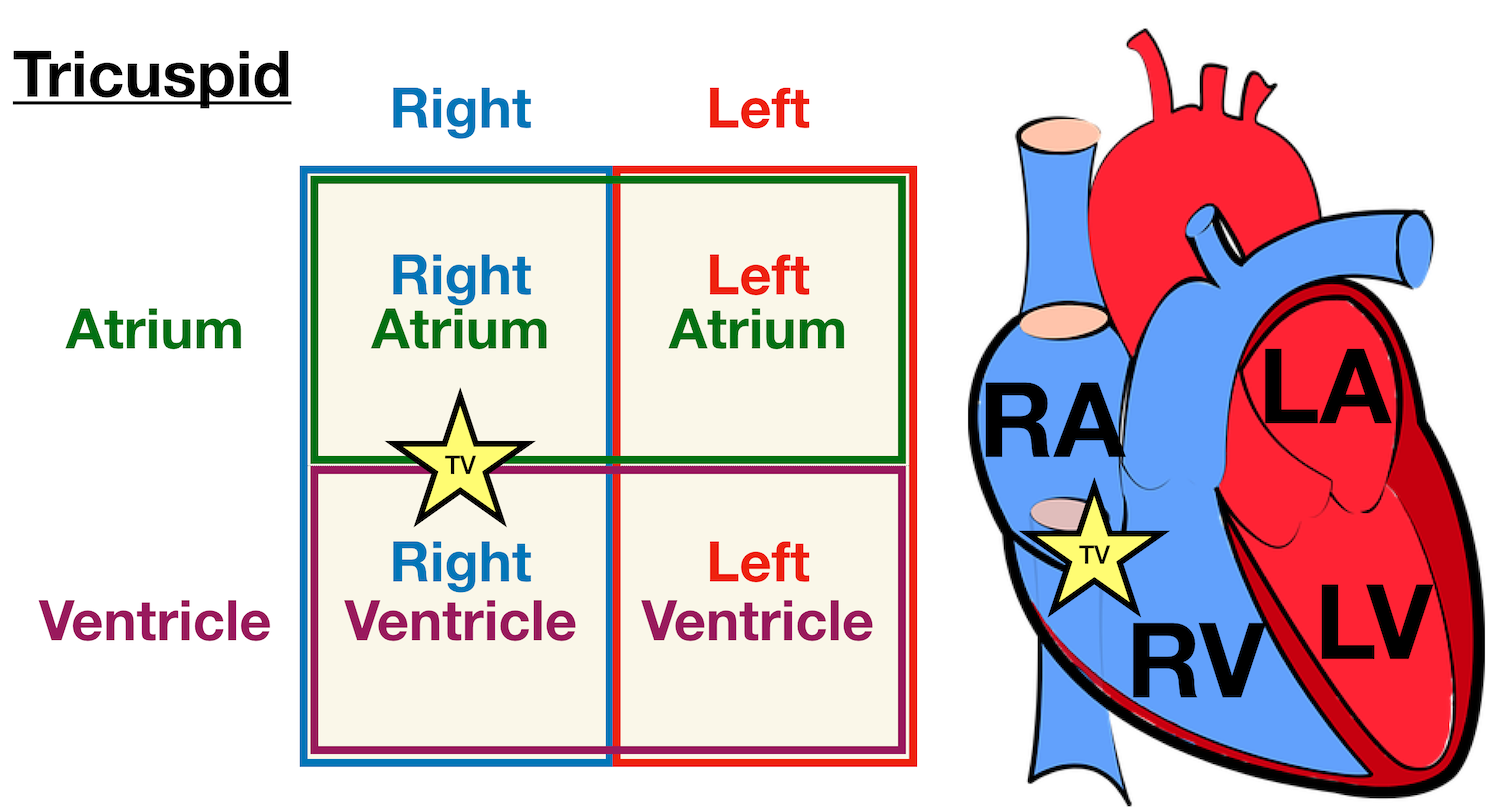
Heart Anatomy: Labeled Diagram, Structures, Blood Flow ... - EZmed There are 4 chambers, labeled 1-4 on the diagram below. To help simplify things, we can convert the heart into a square. We will then divide that square into 4 different boxes which will represent the 4 chambers of the heart. The boxes are numbered to correlate with the labeled chambers on the cartoon diagram. View fullsize
heart | Structure, Function, Diagram, Anatomy, & Facts The heart consists of several layers of a tough muscular wall, the myocardium. A thin layer of tissue, the pericardium, covers the outside, and another layer, the endocardium, lines the inside. The heart cavity is divided down the middle into a right and a left heart, which in turn are subdivided into two chambers.
Heart anatomy: Structure, valves, coronary vessels | Kenhub Heart anatomy. The heart has five surfaces: base (posterior), diaphragmatic (inferior), sternocostal (anterior), and left and right pulmonary surfaces. It also has several margins: right, left, superior, and inferior: The right margin is the small section of the right atrium that extends between the superior and inferior vena cava .
A Labeled Diagram of the Human Heart You Really Need to See The human heart, comprises four chambers: right atrium, left atrium, right ventricle and left ventricle. The two upper chambers are called the left and the right atria, and the two lower chambers are known as the left and the right ventricles. The two atria and ventricles are separated from each other by a muscle wall called 'septum'.
Structure of the Heart | The Franklin Institute The heart consists of four chambers: two atria on the top and two ventricles on the bottom. Looking at the Valentine's Day heart, the two rounded humps at the top are rounded like the top of a lower-case "a." The bottom is shaped like a "v." Feel it working What else is inside your heart?
Label the HEART | Circulatory System Quiz - Quizizz What is number two pointing at in the heart diagram? answer choices Right Atrium Right Ventricle Left Atrium Left Ventricle Question 2 60 seconds Q. What is number one pointing at in the heart diagram? answer choices Right Ventricle Right Pulmonary Vein Superior Franklin Inferior Vena Cava Question 3 60 seconds Q.
Human Heart - Diagram and Anatomy of the Heart - Innerbody The heart contains 4 chambers: the right atrium, left atrium, right ventricle, and left ventricle. The atria are smaller than the ventricles and have thinner, less muscular walls than the ventricles. The atria act as receiving chambers for blood, so they are connected to the veins that carry blood to the heart.
Label the heart — Science Learning Hub In this interactive, you can label parts of the human heart. Drag and drop the text labels onto the boxes next to the diagram. Selecting or hovering over a box will highlight each area in the diagram. pulmonary vein semilunar valve right ventricle right atrium vena cava left atrium pulmonary artery aorta left ventricle Download Exercise Tweet
Anabolic steroid - Wikipedia Most steroid users are not athletes. In the United States, between 1 million and 3 million people (1% of the population) are thought to have used AAS. Studies in the United States have shown that AAS users tend to be mostly middle-class men with a median age of about 25 who are noncompetitive bodybuilders and non-athletes and use the drugs for cosmetic purposes. "
Heart: Anatomy and Function - Cleveland Clinic Your heart walls have three layers: Endocardium: Inner layer. Myocardium: Muscular middle layer. Epicardium: Protective outer layer. The epicardium is one layer of your pericardium. The pericardium is a protective sac that covers your entire heart. It produces fluid to lubricate your heart and keep it from rubbing against other organs.
Sotalol: Uses, Interactions, Mechanism of Action | DrugBank ... Pharmacodynamics. Sotalol is a competitive inhibitor of the rapid potassium channel. 2 This inhibition lengthens the duration of action potentials and the refractory period in the atria and ventricles. 3,4 The inhibition of rapid potassium channels is increases as heart rate decreases, which is why adverse effects like torsades de points is more likely to be seen at lower heart rates. 6 L ...
Heart Labels - Printable or Custom Printed Stickers | Avery.com Use our free specialty shape label templates to easily personalize your heart labels online. Customize one of our free designs or upload your own graphics and then choose the printing option that works best for you. Order your blank heart labels or custom printed heart labels and stickers online and get free shipping on orders of $50 more.
Structure and function of the heart - BBC Bitesize It is located in the middle of the chest and slightly towards the left. The heart is a large muscular pump and is divided into two halves - the right-hand side and the left-hand side. The...
[IGCSE/GCSE] Heart Structure - Memorize In 5 Minutes Or Less! 📘 FREE Comprehensive notes onhttps:// ️Join patreon to access EXCLUSIVE content! ️ Past ...
Heart Diagram with Labels and Detailed Explanation - Collegedunia The heart is located under the ribcage, between the lungs and above the diaphragm. It weighs about 10.5 ounces and is cone shaped in structure. It consists of the following parts: Heart Detailed Diagram. Heart - Chambers There are four chambers of the heart. The upper two chambers are the auricles and the lower two are called ventricles.
Marketing Week | marketing news, opinion, trends and jobs Marketing Week offers the latest marketing news, opinion, trends, jobs and challenges facing the marketing industry.

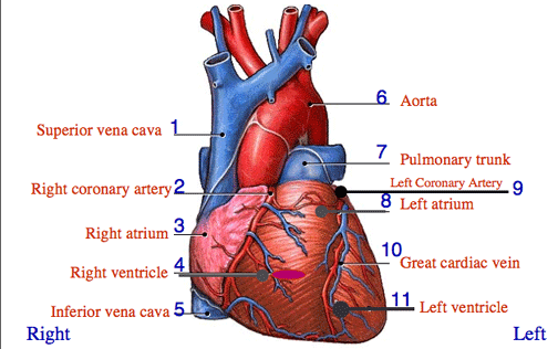



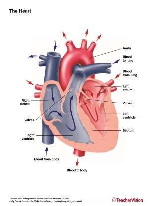









![PDF] Macroscopic Structure and Physiology of the Normal and ...](https://d3i71xaburhd42.cloudfront.net/385136cf4369155490f2aa117e783684aaf8c35c/15-Figure8-1.png)

Post a Comment for "39 heart structure with labels"